The Clinical & Engineering Problem
Dual-energy X-ray absorptiometry (DXA) misses up to 50% of fragility fractures because areal bone mineral density ignores spatial microarchitecture and regional trabecular organization.
The Technical Approach
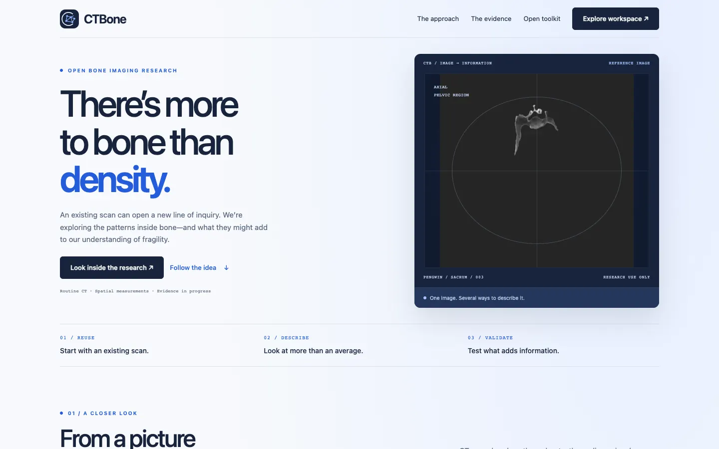
Automates anatomical segmentation of the sacrum and iliac regions on standard abdominal/pelvic CTs, extracting spatial gradient and texture descriptors that capture microstructural heterogeneity.
Engineering Architecture
- Volumetric masking pipeline isolating sacral and bilateral iliac cancellous compartments
- Spatial-texture radiomic feature extraction with Hounsfield unit calibration
- Patient-locked cross-validation with reported uncertainty intervals
- Interactive browser slice viewer demonstrating regional feature extraction
Documented Evidence & Verification
Multi-Region
Segmentation
Sacrum, left iliac and right iliac cancellous compartments
Open Research
Reproducibility
Standardized feature extraction pipeline with JSON fingerprinting
Scientific Status & Regulatory Disclaimers
Research use only. Does not replace DXA or FRAX scores. Clinical superiority to standard bone densitometry requires ongoing prospective multi-center fracture cohort validation.